論文がアクセプトされました(川俣先生・鈴木先生 共著)
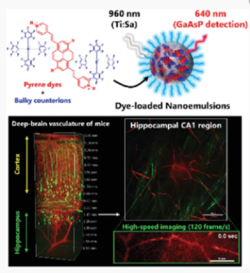
Integrated Fluorescent Nanoprobe Design for High-Speed In Vivo Two-Photon
Microscopic Imaging of Deep-Brain Vasculature in Mice
M. Takezaki, R. Kawakami, S. Onishi, Y. Suzuki, J. Kawamata, T. Imamura, S. Hadano, S. Watanabe, Y. Niko
Dalton Trans., 2021, 31, 2010698
DOI : 10.1002/adfm.202010698
Abstract: High-speed two-photon microscopy can be used to analyze vascular dynamics
in living animals and is essential for the understanding of brain diseases.
Recent advances in fluorescent probes/optical systems have allowed successful
imaging of the hippocampal vasculature in the deep brain of mice (1 mm
from the brain surface) under low-speed conditions (1–2 fps); however,
using high-speed techniques (>30 fps), observation of the deep-brain
vasculature is still challenging. Here, a new nanoemulsion that encapsulates
thousands of red-emissive pyrene dye molecules while maintaining their
high two-photon brightness [1.5 × 10² GM (GM = 10−50 cm4·s·photon−1·molecule−1)
at 960 nm excitation] and delivers a large amount of such pyrene dyes (65
nmol) into the blood vessels of mice is developed. Remarkably, the nanoprobe
is found to exploit the inherent performance of a commonly used Ti:sapphire
excitation laser and a sensitive gallium arsenide phosphide nondescanned
fluorescence detector to the limit, enabling visualization of the brain
vasculature under the cortex region of mice (up to 1.5 mm) under very low-speed
conditions. As a highlight, such a nanoprobe is successfully used to directly
observe the blood flow in the hippocampal CA1 region (1.1 mm) through high-speed
resonant scanning (120 fps).